How it works
Neocision Medical’s patented technology enables the fully automated, transcutaneous capture of a targeted tissue volume under ultrasound guidance having a diameter of up to 30 mm through a small skin incision. The Neocision System is comprised of a reusable Handpiece and removably attachable single-use Wand.
The Handpiece incorporates two independently controlled motor-actuated drive mechanisms. A first drive mechanism advances six leaf members that support a circumscribing wire that thermally cuts through soft tissue as the leaf members are advanced. Once the pre-selected tissue capture diameter is reached by the cutting wire, a second drive motor controls the rate of purse-down of the six leaf members to complete the capture of a nearly spherical tissue volume as seen in the simulated tissue capture video. The automated capture of a targeted tissue volume requires less than about 20 seconds. The selectable video illustrates the simulated capture of a 25 mm diameter tissue volume.
The Handpiece incorporates its own rechargeable battery energy source, control system and operator actuation switches and displays. A toggle switch on the Handpiece allows the operator to select a diameter for tissue capture in the range from 15 mm to 30 mm in 5 mm increments. The Wand is a universal single-use product since the size of the tissue capture is selectable by controls within the Handpiece.
The patented thermal cutting mechanism confines electrical current flow within the cutting wire. As a result, there is no electrical current flow within the body thereby avoiding nerve stimulation and potential nerve injury that can extend outside the zone of local anesthesia causing significant pain to the patient during the procedure. Also, the patented thermal cutting mechanism eliminates the generation of smoke and the release of hazardous volatiles thereby avoiding the need for smoke evacuation required for all monopolar electrosurgical (diathermy) cutting methods.
Using real-time ultrasound guidance, the distal end of the single-use Wand, inserted in the Handpiece, is accurately positioned adjacent to the targeted lesion. A surgically sharp blade located at the distal end of the Wand enables the advancement of the distal end of the Wand into adjacency with the targeted lesion.
Once the distal end of the Wand is in position, six leaf members are automatically deployed to their maximum opening positions and then pursed down, providing a totally enclosing cage around the incised tissue volume. At the completion of the capture, a small marker pellet is implanted into the site of the incised tissue volume for future imaging, if necessary. The contained tissue volume is then withdrawn from the body and provided to a pathologist. The pathologist examines sections of the excised tissue volume to enable characterization of the lesion contained within the captured tissue volume as well as verification of the presence of clear margins between the lesion and the surface of the excised tissue volume.
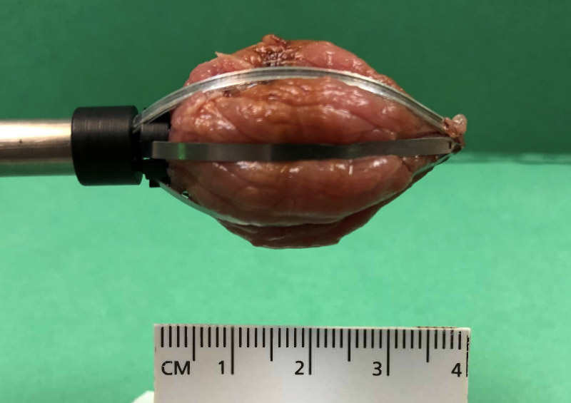
25mm Diameter Tissue Capture Using Wand
Percutaneous Tissue Capture Mechanism
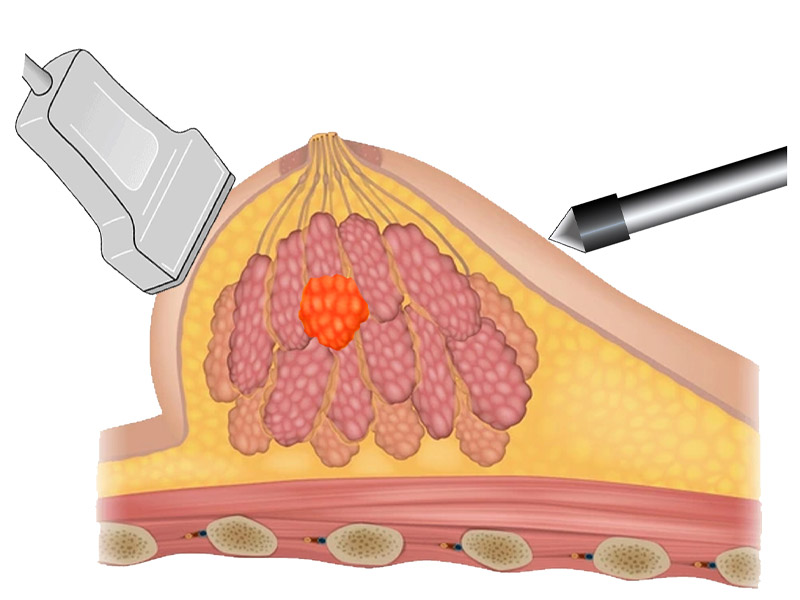
Step 1
Use ultrasound to localize the lesion.
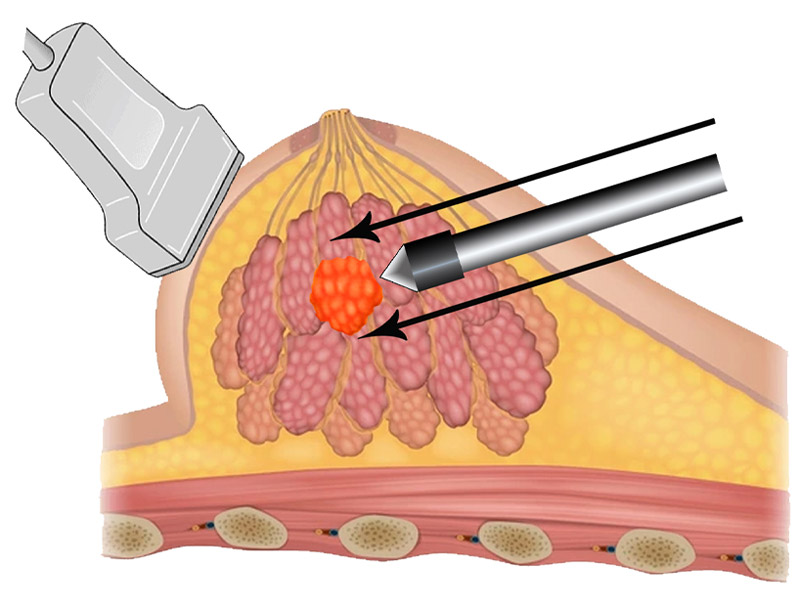
Step 2
Use ultrasound to position wand proximal to lesion.
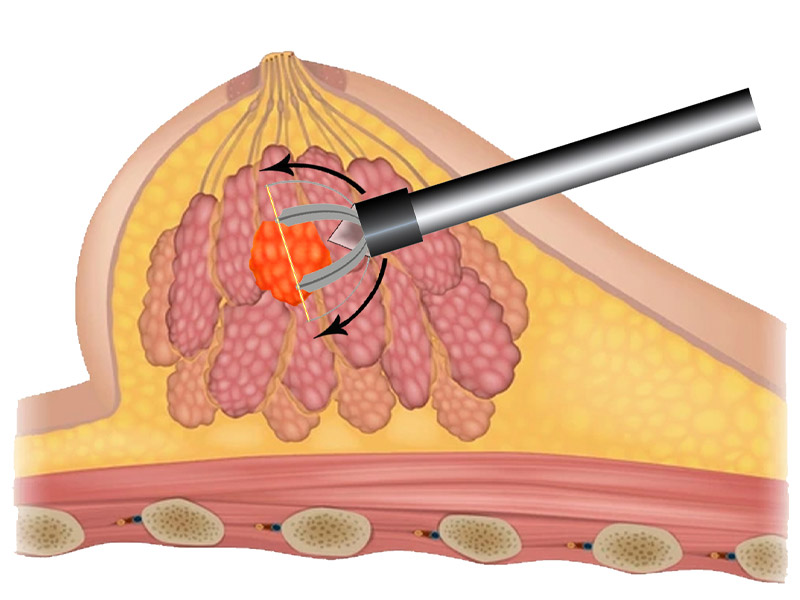
Step 3
Initiate automated capture of lesion.
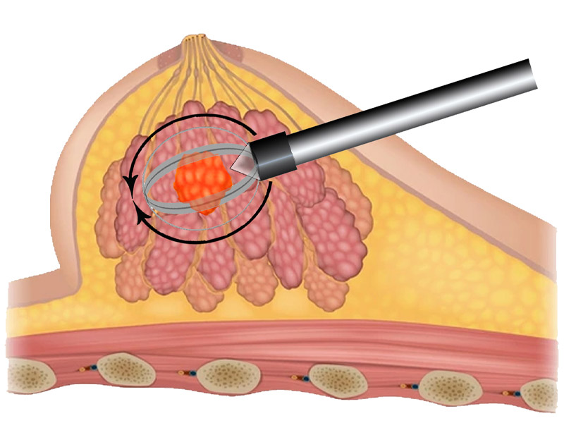
Step 4
Complete automated capture of lesion.
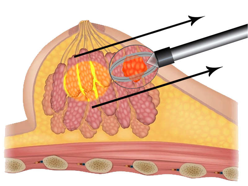
Step 5
Initiate withdrawal of captured lesion.
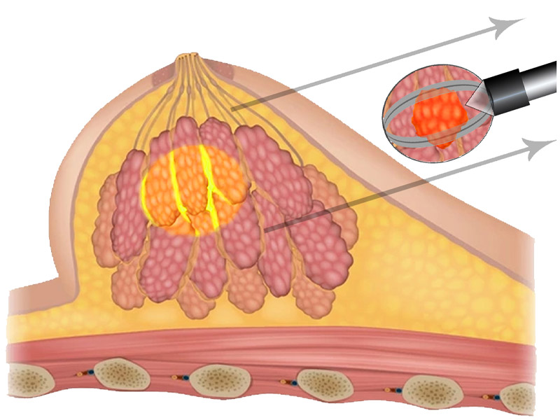
Step 6
Tissue volume containing lesion is submitted for pathology examination.